La Unidad de Imagen del CIMA participa en un proyecto pionero con momias egipcias de animales
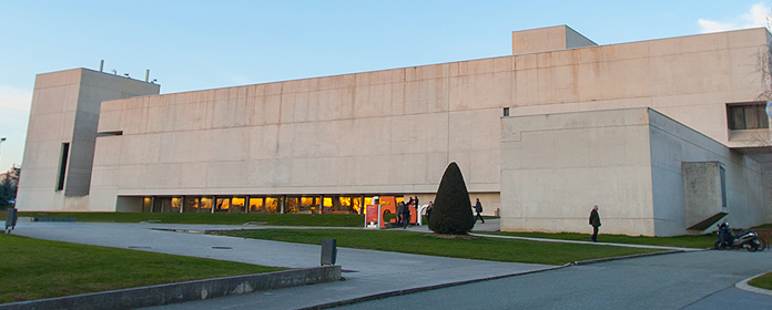
El vestíbulo del edificio nuevo de la Biblioteca de Humanidades acoge hasta el 15 de abril una exposición sobre este estudio
La Unidad de Imagen del Centro de Investigación Médica Aplicada (CIMA) de la Universidad de Navarra ha participado en un proyecto pionero con momias egipcias de animales. Mediante el Micro CT del CIMA y un TAC de la Clínica Universidad de Navarra, los investigadores han estudiado 8 momias de animales de 3.000 años de antigüedad: dos peces de la especie Tilapia nilótica, dos gatos, un halcón, una cabeza de felino, un cocodrilo y un pez mormyrus, que forman parte de colecciones particulares y de museos españoles. "A la vista de las imágenes se aprecia que los embalsamadores situaban a los animales en su posición más natural, los peces por ejemplo están como si nadaran por el agua; el cocodrilo como si reptara, etc.", afirma la egiptóloga Mariluz Mangado.
Se trata de un estudio pionero a nivel mundial, ya que es el primero que se conoce de estas características y que haya sido aplicado a momias no humanas. "Nos ha empujado a colaborar la curiosidad de ver lo que hay dentro de una ‘caja negra', sin abrirla, ya que causaría un daño irreparable, al deshacer el vendaje y provocar la aceleración de la degradación de la muestra", explicó el ingeniero Carlos Ortiz de Solórzano, director de la Unidad de Imagen del CIMA.


