Un análisis de sangre ayudará a prevenir complicaciones clínicas en la miocardiopatía hipertrófica, que afecta a 1 de cada 500 personas
The New England Journal of Medicine has published the results of research carried out by the University of Navarra
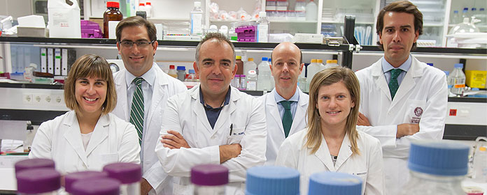
The myocardial fibrosis present in myocardial hypertrophy (MCH) can be detected by biochemistry before thickening of the left ventricle ensues, alongside its clinical complications. This is the main finding outlined in a study published in New England Journal of Medicine, carried out by researchers at the Center for Applied Medical Research (CIMA) and the Clínica Universidad de Navarra, as part of the Cardiovascular Research Network (RECAVA) based at the Instituto de Salud Carlos III, in cooperation with scientists at Harvard University. The team was headed by Dr. Javier Díez, who is in charge of the area of Cardiovascular Sciences at CIMA and Full Professor of Medicine at the University of Navarra.
MCH is the most frequent heart disease of genetic origin. It affects 1 in every 500 people and can lead to sudden death, ventricular arrythmia, ventricular dysfunction and cardiac insufficiency. There are two types of patient with MCH, people with the genetic mutation that causes it, but who do not have thickening of the myocardium (the disease in its pre-clinical phase) and people who have the mutation and also present thickening of the myocardium (the disease in its clinical phase).
Fibrosis of the myocardium is a characteristic lesion in MCH, and one which has a major impact on the development of clinical complications. It had long been thought that the fibrosis developed once the wall of the left ventricle had thickened, but animal studies have shown that the fibrosis is actually present beforehand. Anti-fibrotic treatment is being developed to prevent the occurrence of clinical complications, and so it is vital to detect the miocardial fibrosis at an early stage.
Detection of PICP in blood"The study consists of analyzing various blood biomarkers related to fibrosis in three groups of subjects: healthy people, patients with MCH in its pre-clinical phase, and patients with MCH in its clinical phase", explains Dr. Díez. The researchers have found that the concentration of one of the biomarkers, the so-called C-terminal propeptide of type I procollagen (PICP), is abnormally high in patients with MCH, even in those in whom the disease is in its pre-clinical phase.
This finding has a practical application, because detecting PICP in the blood will make it possible to identify patients with pre-clinical MCH and myocardial fibrosis, and to assess the severity of fibrosis in patients with clinical MCH. This is possible because PICP has been associated with abnormal deposits of collagen fibers in the myocardium. What is more, diagnosis of fibrosis by PICP has been shown to be more sensitive than diagnosis by imaging techniques. Determining PICP levels in people with MCH also makes it possible to design individualized anti-fibrotic treatments to prevent the clinical complications of MCH.
"This study, published in the New England Journal of Medicine, is intended to improve the diagnosis and treatment of this disease, and is a clear example of the applied translational research carried out through the cooperation of scientists from Navarra and Harvard University", explains Dr. Díez.